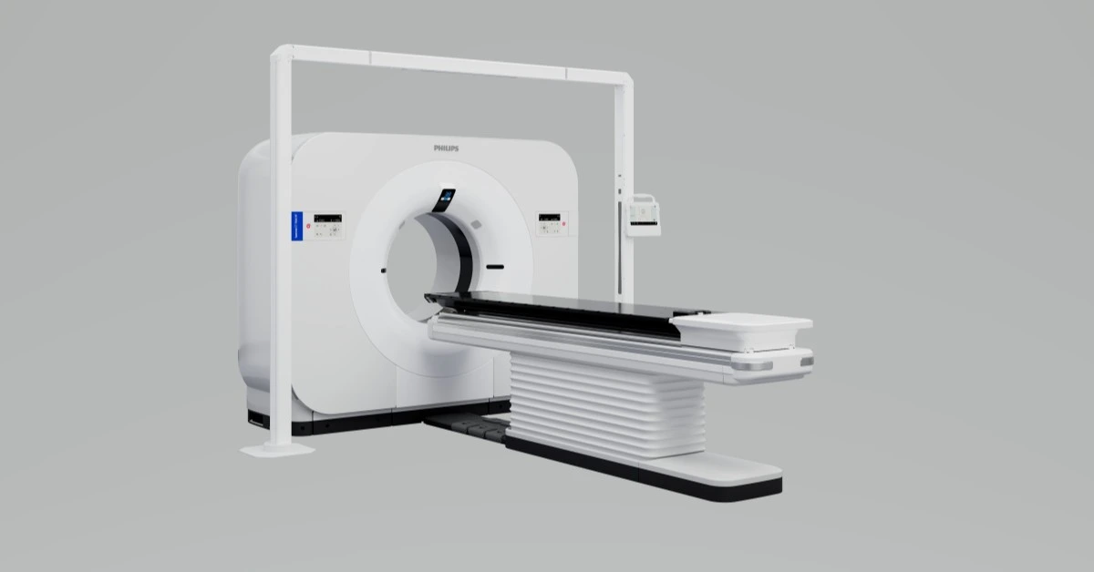
NETHERLANDS – Royal Philips, a global leader in health technology, has achieved FDA 510(k) clearance for its groundbreaking Philips Spectral CT 7500 RT system.
This innovative detector-based spectral CT solution is designed specifically to advance precision in radiation oncology for cancer treatment.
This approval is a major step forward in personalized cancer care, as the system’s unique imaging capabilities allow radiation oncologists to tailor treatment with greater precision than ever before.
The Spectral CT 7500 RT enables healthcare providers to view both conventional and spectral CT images in a single scan, merging advanced tumor visualization and tissue characterization with existing clinical workflows.
This integration empowers radiation oncologists to precisely target radiation therapy to the distinct physiological traits of a patient’s tumor, reducing the potential damage to surrounding healthy tissue and minimizing unwanted side effects.
“With the Spectral CT 7500 RT, radiation oncologists gain access to the critical spectral information needed to enhance tissue characterization,” said Dan Xu, Global Business Leader of CT at Philips.
“This advancement enables wider access to highly personalized and precisely targeted treatment for more patients without adding extra steps to current radiotherapy workflows.”
The Philips Spectral CT 7500 RT is the first radiation therapy CT scanner to incorporate respiratory-gated spectral imaging, combining the benefits of 4D conventional CT with the enhanced visualization and quantification of spectral CT.
This means radiotherapy departments can implement more accurate and effective treatment plans without the added costs of multiple scans.
By reducing the need for additional imaging, Philips’ new system streamlines treatment for a greater number of cancer patients.
According to Philips, the Spectral CT also sets a new standard in accuracy for proton therapy by reducing proton stopping-power ratio (SPR) error by over 50% compared to conventional CT, which helps protect healthy tissue.
The system provides electron density and effective atomic number information in a single scan, automatically generating SPR maps with less than 1% deviation.
This precision significantly improves dose calculations and the accuracy of radiation therapy.
According to Dr. Zhong Su, Physics Director at the Department of Radiation Oncology, University of Arkansas for Medical Sciences, the Spectral CT system has introduced capabilities that conventional CT lacks.
“It can provide electron density and effective atomic number results, which we can convert to the proton stopping-power ratio,” Dr. Su explained.
“Published data shows that the stopping power ratio obtained in this way has fewer uncertainties compared to regular calibration curves, thereby reducing the uncertainty margins during treatment planning.”
Philips has been a pioneering force in medical imaging for over 40 years, contributing to advances that have helped physicians make accurate diagnoses across multiple clinical areas, including oncology, neurology, and cardiology.
The company continues to lead in spectral CT innovation with its “always-on” detector-based technology, now offering transformative solutions in radiation oncology to improve cancer care outcomes worldwide.
For more insights into this new development, Philips will be showcasing the Spectral CT 7500 RT at the RSNA 2024 conference.
XRP HEALTHCARE L.L.C | License Number: 2312867.01 | Dubai | © Copyright 2025 | All Rights Reserved